Calibration curve for an immunoassay.
- Homogeneous immunoassays do not require antibody–bound antigen and free (unbound) antigen to be separated before measurement of the signal.
- Heterogeneous immunoassays require the separation of antibody–bound and free antigen before measurement of the signal.
Antibody production

Figure 2. Structure of diamorphine (heroin) and its major metabolites.
Monoclonal antibodies versus polyclonal antisera
Polyclonal antisera
Immunisation of a rabbit or sheep usually proceeds with 20 to 100 μg of the protein–conjugated hapten, which can be mixed with an adjuvant to stimulate the immune system. This produces an immune response that consists mainly of IgM followed by IgG. This response is further stimulated by additional immunisations of a similar or lower dose at regular intervals – typically every 3 to 4 weeks for 20 weeks or longer. Test bleed samples of approximately 1 to 2 mL are taken at 10 to 14 days post–injection, at which point the immune response (and therefore antibody titre in the serum) is at its highest. After analysis of the test bleeds, a larger antiserum sample can be drawn. This can be as much as several hundred millilitres when using larger host animals (e.g. sheep). Antisera yield can vary from hundreds to many thousands of tests per millilitre depending on the success of the immunisation programme and the type of assay in which they are employed. It is not the absolute volume of serum that is important, but rather the amount and quality of antibodies contained therein. Using standard separation methods, such as ammonium sulfate precipitation, Protein A separation and/or ion–exchange chromatography, antisera can be purified for further use. Affinity chromatography can be beneficial in certain circumstances and involves passing the antiserum over a column that contains an immobilised form of the drug of interest. In this way it is possible to fractionate a complex polyclonal antiserum. In some cases purification is not necessary and the antiserum (i.e. the serum itself) can be used with simple dilution.Monoclonal antibodies
Monoclonal antibodies offer the advantage of a continuous supply of antibodies with the same characteristics, so once a good antibody is selected it can be used indefinitely. After immunisation and successful test bleeds, monoclonal antibodies are made by the fusion of mouse lymphocytes (or lymphocytes from other species) from the spleen with myeloma cells. The resultant hybridoma cells are separated by limiting dilution to give single cells that secrete single monoclonal antibodies. This technique was first described in 1975 and is now in routine use (Kohler and Milstein 1975). Monoclonal antibodies generally have less affinity than polyclonal equivalents, which can lead to less sensitive assays. Monoclonal antibodies are not more specific than polyclonal antisera, but once a specific antibody is selected, the cell line can be stored and the antibody produced indefinitely. Note that in drug testing, it is possible for an antibody to be too specific, as it may be desirable to have broad cross–reactivity to a drug family (such as benzodiazepines) or to a single drug and its metabolites (such as buprenorphine). Monoclonal antibodies offer the advantages of purity and homology, which is useful for circumstances in which the antibody is being labelled or conjugated as part of the immunoassay set up – for example when being labelled with an enzyme or coated with colloidal gold. Other molecular biology and recombinant techniques, such as phage display (Chiswell and McCafferty 1992), in which the genetic code is harnessed to produce antibodies give an exciting additional source of this important immunoassay component.Antibody dilution curves
Once an immunisation is underway, the quality of the antiserum is assessed by means of an antiserum dilution curve. This demonstrates the binding of the antibody to the target drug and is an indication of the antibody’s affinity for the antigen. Using a heterogeneous enzyme immunoassay (EIA) as an example, a test bleed of rabbit anti–cotinine was coated onto a microplate at different dilutions in bicarbonate buffer (pH 9) using incubation overnight at room temperature. The resultant binding to different titres of horseradish peroxidase–labelled cotinine. Dilutions of the enzyme–drug conjugate for this type of experiment can be made in simple phosphate buffer (pH 7.4) with a small amount of protein (e.g. BSA at 0.05% w/v) to prevent non–specific binding (NSB; i.e. non–specific binding) of materials to the assay tube. The hook (or apparent peak) at high antiserum concentrations is caused by a combination of steric hindrance and saturation of coating antibody to the microplate. Once the concentration of antibody (titre) that produces the desired response is selected, it is necessary to check that the antibody also responds as required with the target drug (i.e. that the target drug successfully competes with the labelled drug for binding to the antibody). The improved performance with subsequent test bleeds for samples taken from a rabbit immunised with a buprenorphine-protein immunogen. It is possible to gain useful information by combining both the experiments described above and analysing both a positive and negative sample at each antibody dilution. This way, binding and displacement can be seen at each antibody titre. Careful titering of the labelled drug derivative and antibody dilution can improve the assay characteristics, and the assay can be optimised further by the addition of other proteins, surfactants and stabilisers to the assay buffer. An immunisation programme usually involves the injection of between three and six animals with the same antigen. If suitable antibodies are not produced after several immunisations, it may be necessary to start the programme again with different animals and possibly a different immunogen.Analytical specificity
Heterogeneous immunoassays

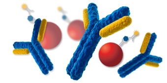
Basics of an immunoassay.
Enzyme immunoassays
Antibody–capture systems
The system uses anti–drug antibodies coated onto a microplate well by passive absorption. A microplate is a tray of 96 wells (each having a volume of approximately 400 μL) that are generally arranged as 12 × 8–well strips. The coating can be achieved by simply adding a solution of the antiserum or purified antibody in a bicarbonate buffer at pH 9 using high–precision pipettes. A suitable incubation period (e.g. overnight at room temperature) allows the antibodies to bind to the plastic microplate well. Following this incubation, the solution is washed from the solid phase and dried to leave an antibody–coated well. In some cases, a secondary coating of a non–relevant protein is performed to increase stability of the solid–phase antibody and to prevent NSB of assay components to the plastic well of the microplate. This process forms part of the reagent manufacturing process of commercial kits provided as a dry microplate with the antibody pre–coated. In running the assay, a sample (10 μL to 50 μL) is added to the microplate followed by a buffered solution (typically 100 μL) that contains a fixed amount of drug labelled with an enzyme such as horseradish peroxidase or alkaline phosphatase. The plate is then left for sufficient time to allow the horseradish peroxidase–labelled drug and any drug present in the sample to compete for binding to the solid–phase antibody. This is referred to as the incubation period. In the absence of drug, the maximum binding of the enzyme label occurs. Increased amounts of drug in the sample result in decreased amounts of antibody–bound enzyme label. After the incubation period, the microplate is washed with a suitable buffer (the separation step) to remove all traces of unbound enzyme label. Antibody–bound enzyme conjugate (and antibody–bound drug) is left immobilised to the wall of the antibody–coated microplate. After the wash step, a substrate reagent is added to the microplate well and left to develop colour. The substrate of choice for horseradish peroxidase is dilute hydrogen peroxide with tetramethylbenzidine (TMB) as chromagen, which is available as a liquid–stable, ready–to–use product. TMB provides a blue coloured signal that can be measured at 630 nm using a standard laboratory microplate reader. In routine use, the colour development is stopped, after it has developed over a 20 to 30 min incubation period, by the addition of 1 M dilute sulfuric acid. This causes a shift in absorbance that is read at 450 nm. The whole assay process described here can be fully automated or performed with the simplest of laboratory equipment. The amount of enzyme–labelled drug bound to the solid–phase antibody, and therefore available for colour development, is inversely proportional to the amount of drug in the specimen. The colour produced at the final stage of the EIA is therefore inversely proportional to the amount of drug in the specimen, which gives a calibration curve similar to that found with RIA .Enzyme–linked immunosorbent assay, antibody labelled systems
Competitive ELISA procedures are similar to the antibody–coated microplate EIA described above. The difference is that the anti–drug antibody is enzyme labelled rather than the drug. A drug derivative is coated onto the plastic well, which serves as a way to separate bound and free fractions. The drug is conjugated to a protein using a process similar to that used to prepare an immunogen. This must be a different protein from the one used as the carrier protein to make the antibody in order to prevent binding of anti–protein antibodies made during the immunisation process. The protein–drug conjugate can be coated to the microplate well in the same manner used for coating antibodies in EIA (see above). During the assay, using similar procedures to those described for EIA, competition occurs between the drug in the sample and the immobilised drug for binding to an enzyme labelled anti–drug antibody. Following incubation, a wash step is used to separate bound and free fractions, and leave labelled antibody immobilised to the solid–phase drug derivative on the microplate well wall. The amount of labelled antibody that is bound, and responsible for the signal generation, is inversely proportional to concentration of drug in the sample.
The basic scheme for an ELISA assay. Note the wash between steps. This is the distinctive feature of a heterogeneous assay.

The scheme shows a typical competitive solid-phase immunoassay commonly used to detect drugs of abuse. It is a heterogeneous format using washing steps to remove materials that did not bind immunologically. Signal is inversely proportional to the concentration of free drug in the sample.
Radioimmunoassay
Chemiluminescence immunoassays
Fluorescent labels
Lateral flow methods
There is a growing trend in the immunoassay sector to develop point–of–care tests. The Syva Emit system (see Homogeneous methods) on small analysers, such as the ETS, has allowed drug testing outside the laboratory for many years (Centofanti 1994). The use of inexpensive, easy–to–use single disposable cartridges or slides for a variety of drugs has accelerated this trend.Homogeneous immunoassays

EMIT assay scheme; signal is directly proportional to the concentration of analyte.
CEDIA, cloned enzyme donor immunoassay
Fluorescence polarisation immunoassay
Micro–particle methods
Automation of immunoassay
Analysis of alternative samples to urine
Quality control, calibration, standardisation and curve fitting

Example to illustrate how different curve–fitting programs can influence the results of an immunoassay.
Troubleshooting

Flow chart to assist with troubleshooting problems encountered with immunoassays.
Within–assay imprecision
All immunoassays have an inherent imprecision and for this reason it has been common practice to perform replicate analyses. For a particular immunoassay, the agreement between replicates is a consistent feature that can be assessed for each assay. Poor replication is easily identifiable and is often associated with a pattern. Those laboratories that only analyse singletons lack this early warning system.Batch–to–batch imprecision
- Level of drug beyond the calibration range (above or below) of the assay.
- Matrix effect through the use of an animal–based control that behaves differently to the material (e.g. human) being tested.
- Use of a control in one matrix in an immunoassay designed for another matrix (e.g. a urine control used in a plasma assay).
- Inadequate reconstitution of control material and deterioration of the control material through long–term storage or repeated freeze–thaw cycles.
External quality assessment performance
Sample adulteration
Références
| ↑1 | Clarke's Analysis of Drugs and Poisons |
|---|
Last modified: 5 December, 2016


